Benefits of Early Detection and Treatment for Glaucoma
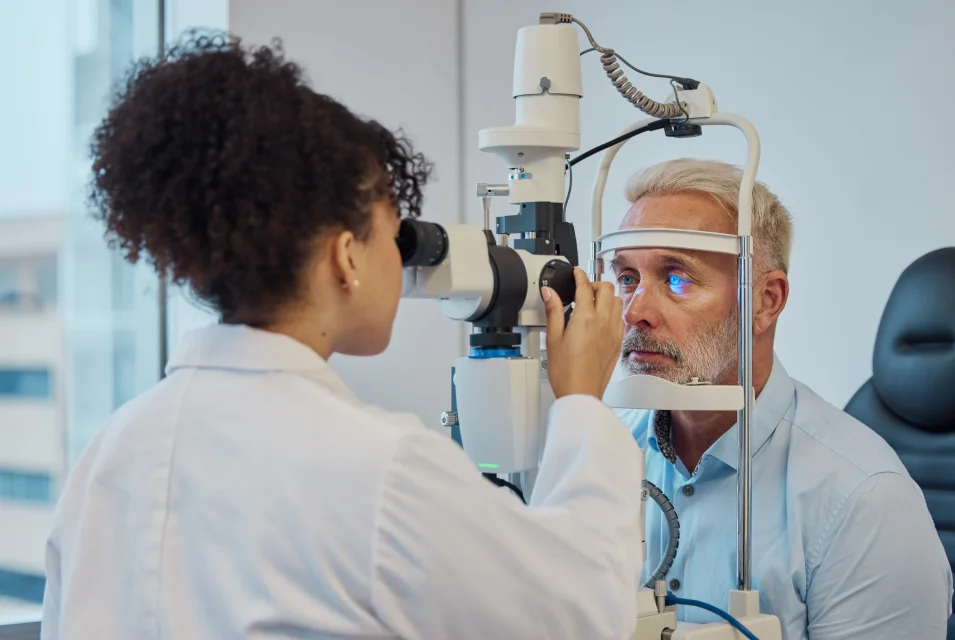
Fundus photography involves photographing the rear of an eye, also known as the fundus. Specialized fundus cameras consisting of an intricate microscope attached to a flash-enabled camera are used in fundus photography. The main structures that can be visualized in a fundus photo are the central and peripheral retina, the optic disc, and the macula. Fundus photography is used by optometrists, ophthalmologists, orthoptists, and other trained medical professionals to monitor the progression of certain eye conditions and diseases. In patients with diabetes mellitus, regular fundus screening examinations are important.
Our Dedicated Specialists


Dr. Amila De Alwis
MBBS,MD OPTHALMOLOGY
FRCS Ophth (Glasgow)
SLMC 17126


Dr. Abhaya Jayasekara
MBBS,MD OPTHALMOLOGY
FRCS Ophth (Glasgow)
SLMC 17126


Dr. Chaya Samarasinghe
MBBS,MD OPTHALMOLOGY
FRCS Ophth (Glasgow)
SLMC 17126


Dr. Duleepa Baranage
MBBS,MD OPTHALMOLOGY
FRCS Ophth (Glasgow)
SLMC 17126


Dr. Chaya Samarasinghe
MBBS,MD OPTHALMOLOGY
FRCS Ophth (Glasgow)
SLMC 17126


Dr. Amila De Alwis
MBBS,MD OPTHALMOLOGY
FRCS Ophth (Glasgow)
SLMC 17126


Call Us on our hotline